



Sonwane Suchitra Shivaji Rao and K. Gangadhara Rao
Department of Computer Science Engineering, Acharya Nagarjuna University
Received: June 17, 2026
Accepted: August 29, 2026
Publication Date: October 26, 2026
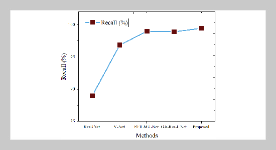
Recall comparison graph
Copyright The Author(s). This is an open access article distributed under the terms of the Creative Commons Attribution License (CC BY 4.0), which permits unrestricted use, distribution, and reproduction in any medium, provided the original author and source are cited.
Download Citation: BibTeX | http://dx.doi.org/10.6180/jase.202701_36.052
Medical analytics based on multimodal intelligence are essential to contemporary healthcare, especially in the diagnosis and detection of liver cancer. However, low contrast, varied textures, and irregular tumor forms in medical imaging continue to make precise liver tumor segmentation difficult. These intricate features are frequently difficult for traditional intensity-based and classical deep learning techniques to adequately capture. This study suggests a unique Inspired Quantum-Classical Sequential Neural Network (IQCSNN) for accurate liver tumor picture segmentation in order to overcome these drawbacks. The suggested method is new because it combines classical sequential neural networks with quantum computational concepts to improve segmentation accuracy and feature representation. To enhance picture quality, preprocessing methods such as normalization, noise reduction, and standardization are used to liver CT images obtained via Kaggle. Following preprocessing, the data is sent into the IQCSNN model, which uses quantum-classical feature extraction techniques to identify both local and global patterns. The Inspired quantum layer enhances the ability to distinguish between tumor and non-tumor tissues, while the sequential neural network component facilitates efficient learning of spatial connections. The usefulness and potential of the suggested strategy for advanced medical image analysis are highlighted by experimental findings, which show that it produces better segmentation performance than traditional methods.
Keywords: Quantum computing; Liver Tumor Segmentation; Sequential neural network; Normalization; Multimodal Feature Extraction
- [1] P. Bilic, P. Christ, H. B. Li, E. Vorontsov, A. Ben-Cohen, G. Kaissis, and B. Menze, (2023) “The Liver Tumor Segmentation Benchmark (LiTS)” Medical Image Analysis 84: 102680. DOI: 10.1016/j.media.2022.102680.
- [2] J. Wang, Y. Peng, S. Jing, L. Han, T. Li, and J. Luo, (2023) “A Deep-Learning Approach for Segmentation of Liver Tumors in Magnetic Resonance Imaging Using UNet++” BMC Cancer 23(1): 1060. DOI: 10.1186/s12885-023-11432-х.
- [3] G. Chen, Z. Li, J. Wang, J. Wang, S. Du, J. Zhou, and Y. Zhou, (2023) “An Improved 3D KiU-Net for Segmentation of Liver Tumor” Computers in Biology and Medicine 160: 107006. DOI: 10.1016/j.compbiomed.2023.107006.
- [4] S. Di, Y. Q. Zhao, M. Liao, F. Zhang, and X. Li, (2022) “TD-Net: A Inspired End-to-End Network for Automatic Liver Tumor Segmentation from CT Images” IEEE Journal of Biomedical and Health Informatics 27(3): 1163-1172. DOI: 10.1109/JBHI.2022.3181974.
- [5] J. Amin, M. A. Anjum, M. Sharif, S. Kadry, and R. G. Crespo, (2023) “Visual Geometry Group Based on U-Shaped Model for Liver/Liver Tumor Segmentation” IEEE Latin America Transactions 21(4): 557-564. DOI: 10.1109/TLA.2023.10128927.
- [6] Y. Chen, C. Zheng, W. Zhang, H. Lin, W. Chen, G. Zhang, and F. Wu, (2023) “MS-FANet: Multi-Scale Feature Attention Network for Liver Tumor Segmentation” Computers in Biology and Medicine 163: 107208. DOI: 10.1016/j.compbiomed.2023.107208.
- [7] C. Zhang, J. Lu, Q. Hua, C. Li, and P. Wang, (2022) “SAA-Net: U-Shaped Network with Scale-Axis-Attention for Liver Tumor Segmentation” Biomedical Signal Processing and Control 73: 103460. DOI: 10.1016/j.bspc.2021.103460.
- [8] J. Li, K. Liu, Y. Hu, H. Zhang, A. A. Heidari, H. Chen, and H. Elmannai, (2023) “Eres-UNet++: Liver CT Image Segmentation Based on High-Efficiency Channel Attention and Res-UNet++” Computers in Biology and Medicine 158: 106501. DOI: 10.1016/j.compbiomed.2022.106501.
- [9] X. Wang, S. Wang, Z. Zhang, X. Yin, T. Wang, and N. Li, (2023) “CPAD-Net: Contextual Parallel Attention and Dilated Network for Liver Tumor Segmentation” Biomedical Signal Processing and Control 79: 104258. DOI: 10.1016/j.bspc.2022.104258.
- [10] S. Di, Y. Zhao, M. Liao, Z. Yang, and Y. Zeng, (2022) “Automatic Liver Tumor Segmentation from CT Images Using Hierarchical Iterative Superpixels and Local Statistical Features” Expert Systems with Applications 203: 117347. DOI: 10.1016/j.eswa.2022.117347.
- [11] F. Lyu, M. Ye, A. J. Ma, T. C. F. Yip, G. L. H. Wong, and P. C. Yuen, (2022) “Learning from Synthetic CT Images via Test-Time Training for Liver Tumor Segmentation” IEEE Transactions on Medical Imaging 41(9): 2510-2520. DOI: 10.1109/TMI.2022.3166230.
- [12] X. Wang, X. Zhang, G. Wang, Y. Zhang, X. Shi, H. Dai, and X. Meng, (2022) “TransFusionNet: Semantic and Spatial Features Fusion Framework for Liver Tumor and Vessel Segmentation under JetsonTX2” IEEE Journal of Biomedical and Health Informatics 27(3): 1173-1184. DOI: 10.1109/JBHI.2022.3207233.
- [13] M. Rela, N. R. Suryakari, and R. R. Patil, (2023) “A Diagnosis System by U-Net and Deep Neural Network Enabled with Optimal Feature Selection for Liver Tumor Detection Using CT Images” Multimedia Tools and Applications 82(3): 3185-3227. DOI: 10.1007/s11042-022-13381-2.
- [14] D. T. Kushnure, S. Tyagi, and S. N. Talbar, (2023) “LiM-Net: Lightweight Multi-Level Multi-Scale Network with Deep Residual Learning for Automatic Liver Segmentation in CT Images” Biomedical Signal Processing and Control 80: 104305. DOI: 10.1016/j.bspc.2022.104305.
- [15] F. Zhan, W. Wang, Q. Chen, Y. Guo, L. He, and L. Wang, (2023) “Three-Direction Fusion for Accurate Volumetric Liver and Tumor Segmentation” IEEE Journal of Biomedical and Health Informatics 28(4): 2175-2186. DOI: 10.1109/JBHI.2023.3344392.
- [16] K. N. Wang, S. X. Li, Z. Bu, F. X. Zhao, G. Q. Zhou, S. J. Zhou, and Y. Chen, (2024) “SBCNet: Scale and Boundary Context Attention Dual-Branch Network for Liver Tumor Segmentation” IEEE Journal of Biomedical and Health Informatics 28(5): 2854-2865. DOI: 10.1109/JBHI.2024.3370864.
- [17] H. Rahman, N. B. Aoun, T. F. N. Bukht, S. Ahmad, R. Tadeusiewicz, P. Pławiak, and M. Hammad, (2025) “Automatic Liver Tumor Segmentation of CT and MRI Volumes Using Ensemble ResUNet-Inception V4 Model” Information Sciences 704: 121966. DOI: 10.1016/j.ins.2025.121966.
- [18] Y. Ni, G. Chen, Z. Feng, H. Cui, D. Metaxas, S. Zhang, and W. Zhu, (2024) “DA-Tran: Multiphase Liver Tumor Segmentation with a Domain-Adaptive Transformer Network” Pattern Recognition 149: 110233. DOI: 10.1016/j.patcog.2023.110233.
- [19] D. Cheng, Z. Zhou, and J. Zhang, (2024) “EG-UNETR: An Edge-Guided Liver Tumor Segmentation Network Based on Cross-Level Interactive Transformer” Biomedical Signal Processing and Control 97: 106739. DOI: 10.1016/j.bspc.2024.106739.
- [20] H. Liu, J. Yang, C. Jiang, S. He, Y. Fu, S. Zhang, and W. Ji, (2024) “S2DA-Net: Spatial and Spectral-Learning Double-Branch Aggregation Network for Liver Tumor Segmentation in CT Images” Computers in Biology and Medicine 174: 108400. DOI: 10.1016/j.compbiomed.2024.108400.
- [21] L. Zhang, W. Zhang, and Z. Xu, (2025) “Automatic Liver Tumor Segmentation Based on Improved YOLO-05 and B-Spline Level Set” Cluster Computing 28(3): 214. DOI: 10.1007/s10586-024-04918-1.
- [22] R. Kılıç, A. Yalçın, F. Alper, E. A. Oral, and I. Y. Ozbek, (2025) “Two-Stage Automatic Liver Classification System Based on Deep Learning Approach Using CT Images” Journal of Imaging Informatics in Medicine: 1-23. DOI: 10.1007/s10278-025-01480-z.
- [23] K. Trivedi, (2025) “Optimization Approaches Using Machine Learning Techniques to Preidentification of Liver Cancer” NanoMind: Exploring Synergies in Nanotechnology and Machine Learning: 151-195. DOI: 10.1007/978-3-031-77296-2_8.
- [24] G. Aksholak, A. Bedelbayev, R. Magazov, and K. Kaplan, (2026) “Transformer Tokenization Strategies for Network Intrusion Detection: Addressing Class Imbalance Through Architecture Optimization” Computers 15(2): 75. DOI: 10.3390/computers15020075.
- [25] R. Jin, H. Y. Tang, Q. Yang, and W. Chen, (2025) “LA-ResUNet: Attention-Based Network for Longitudinal Liver Tumor Segmentation from CT Images” Computerized Medical Imaging and Graphics 123: 102536. DOI: 10.1016/j.compmedimag.2025.102536.
- [26] A. B. Slama, H. Sahli, Y. Amri, and S. Labidi, (2025) “V-NET-VGG16: Inspired Deep Learning Architecture for Optimal Segmentation and Classification of Multi-Differentiated Liver Tumors” Intelligence-Based Medicine 11: 100210. DOI: 10.1016/j.ibmed.2025.100210.
- [27] J. Zhao and S. Li, (2025) “Uncertainty-Guided and Cross-Modality Attention Network for Liver Tumor Segmentation and Quantification via Integrating Dynamic MRI” Knowledge-Based Systems 310: 113021. DOI: 10.1016/j.knosys.2025.113021.
- [28] P. Sivanagaraju, S. V. Ramana, and P. P. Reddy, (2025) “An Advanced Transformer Framework for Liver Tumor Segmentation Using MRI Images” Biomedical Signal Processing and Control 107: 107808. DOI: 10.1016/j.bspc.2025.107808.
- [29] G. N. Yashaswini, R. V. Manjunath, B. Shubha, P. Prabha, N. Aishwarya, and H. M. Manu, (2025) “Deep Learning Technique for Automatic Liver and Liver Tumor Segmentation in CT Images” Journal of Liver Transplantation 17: 100251. DOI: 10.1016/j.liver.2024.100251.
- [30] L. W. Tsai, A. Agrawal, P. Dash, S. Panda, Y. W. Huang, S. Saxena, and C. M. Shih, (2025) “RFILM U-Net: Radiomic Feature-Integrated Linear Modulation Network for Precise Liver Tumor Segmentation” Journal of Medical and Biological Engineering 45(2): 177-186. DOI: 10.1007/s40846-025-00938-3.
- [31] J. K. Appati, N. A. Azuponga, L. M. Boante, and J. A. Mensah, (2024) “A Multiattention ResUNet and Modified U-Net Architecture for Liver Tumor Segmentation” Applied Computational Intelligence and Soft Computing 2024(1): 8365349. DOI: 10.1155/2024/8365349.
- [32] M. Reyad, A. M. Sarhan, and M. Arafa, (2024) “Architecture Optimization for Inspired Deep Residual Networks in Liver Tumor Segmentation Using a GA” International Journal of Computational Intelligence Systems 17(1): 209. DOI: 10.1007/s44196-024-00542-4.
- [33] Y. Challagundla, T. S. C. Tunuguntla, S. G. Tunuguntla, and N. Devarakonda, (2025) “Convolutional Neural Network-Based Classifiers for Liver Tumor Detection Using Computed Tomography Scans” Innovations in Systems and Software Engineering 21(2): 533-539. DOI: 10.1007/s11334-023-00547-w.
- [34] C. Bai, W. Shi, M. Zhang, and H. Zhao, (2026) “Crack-DualMamba: A Lightweight Dual-Stream Mamba with Novel Focal Dice Balanced Loss for Vehicle-Based Road Crack Segmentation” Advanced Engineering Informatics 71: 104427. DOI: 10.1016/j.aei.2026.104427.
- [35] J. Zhang, C. Bai, W. Pan, and J. Guo, (2026) “Virtual-Force Based Visual Servo for Multiple Peg-in-Hole Assembly With Tightly Coupled Multi-Manipulator” IEEE Robotics and Automation Letters 11(3): 2586-2593. DOI: 10.1109/LRA.2026.3653374.
- [36] A. B. Sawad and M. Binsawad, (2026) “Radiomics-Driven Inspired Deep Learning for MRI-Based Prediction of Glioma Grade and 1p/19q Codeletion” Tomography 12(2): 25. DOI: 10.3390/tomography12020025.
- [37] B. A. Alqaralleh, M. Z. Alksasbeh, A. Kulakli, and A. I. Zreikat, (2026) “An Intelligent Healthcare Framework for Hepatocellular Carcinoma Diagnosis Based on Aggregated Learners from Biomedical Data Utilizing Explainable Artificial Intelligence” Scientific Reports 16: 9357. DOI: 10.1038/s41598-026-39871-z.
- [38] E. Lanza, A. Ammirabile, A. Vanzulli, C. Lisi, A. S. Perera, A. M. A. Lucia, and A. Laghi, (2026) “3D Region-Growing nnU-Net Improves Pulmonary Embolism Detection on CTPA: A Dual-Cohort Validation Study” European Radiology Experimental 10(1): 23. DOI: 10.1186/s41747-026-00693-3.
- [39] P. Sun, J. Yu, Q. Gu, L. Zhang, Y. Sun, Q. Wang, and J. Zhu, (2026) “Clinically Oriented Automatic 2D Liver Tumor Segmentation: LCMambaNet with a State-Space Model and Liver Cancer-Specific Attention” Frontiers in Oncology 16: 1676424. DOI: 10.3389/fonc.2026.1676424.
- [40] A. I. Abian, R. K. Debnath, M. A. Rahman, M. A. K. Raiaan, M. R. Islam, A. Karim, and S. Azam, (2026) “HANS-Net: Hyperbolic Convolution and Adaptive Temporal Attention for Accurate and Generalizable Liver and Tumor Segmentation in CT Imaging” IEEE Transactions on Radiation and Plasma Medical Sciences: DOI: 10.1109/TRPMS.2026.3654628.